10+ Bence Jones Protein Tips For Accurate Urine Analysis

The presence of Bence Jones proteins in urine is a crucial diagnostic marker for multiple myeloma and other plasma cell dyscrasias. These proteins are composed of light chains of immunoglobulins and can be detected through various laboratory tests. Accurate urine analysis is essential for the diagnosis and monitoring of these conditions. Here, we will discuss 10+ tips for accurate urine analysis to detect Bence Jones proteins, focusing on the latest techniques, troubleshooting, and interpretation of results.
Introduction to Bence Jones Proteins

Bence Jones proteins are abnormal proteins found in the urine of patients with multiple myeloma, Waldenström’s macroglobulinemia, and other lymphoproliferative disorders. These proteins are produced by neoplastic plasma cells and can be either kappa or lambda light chains. The detection of Bence Jones proteins in urine is a critical diagnostic criterion for multiple myeloma, and their quantification can help monitor disease progression and response to treatment.
Collection and Preparation of Urine Samples
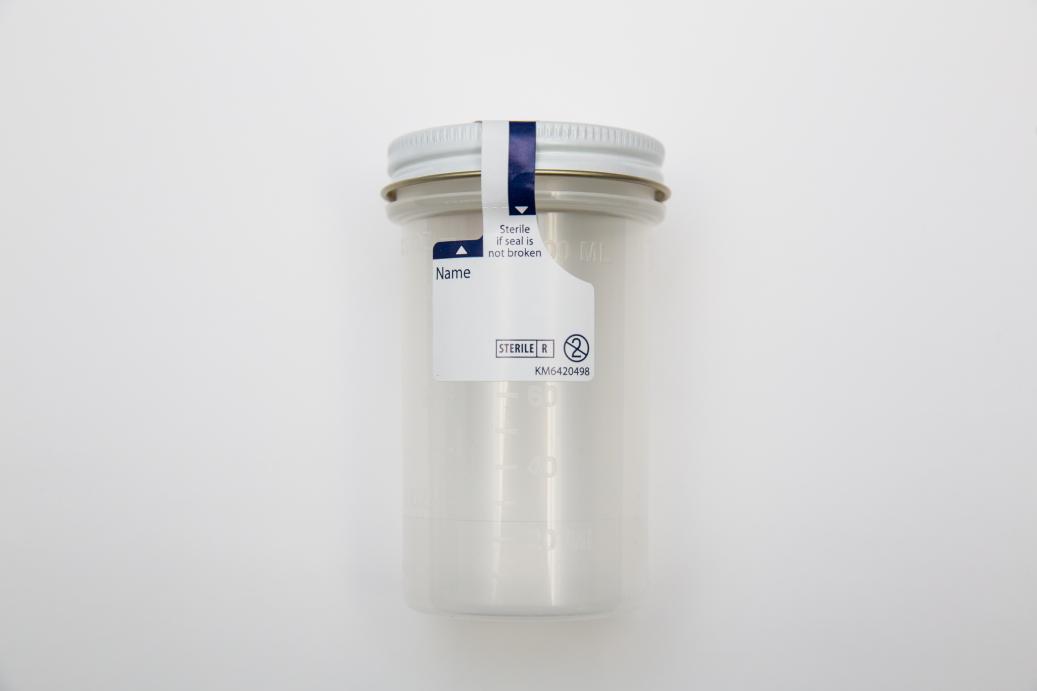
The collection and preparation of urine samples are critical steps in the detection of Bence Jones proteins. Proper collection techniques should be followed to avoid contamination and ensure accurate results. A random urine sample is often sufficient for screening, but a 24-hour urine collection may be necessary for quantification and monitoring. It is essential to store urine samples at 4°C to prevent protein degradation and to analyze them within 24 hours of collection.
| Urine Sample Type | Description |
|---|---|
| Random Urine Sample | Suitable for initial screening |
| 24-hour Urine Collection | Necessary for quantification and monitoring |

Detection Methods for Bence Jones Proteins

Several methods are available for detecting Bence Jones proteins in urine, including electrophoresis, immunofixation electrophoresis, and immunoturbidimetry. Each method has its advantages and limitations, and the choice of method depends on the laboratory’s equipment and expertise. Electrophoresis is a widely used technique that separates proteins based on their size and charge, while immunofixation electrophoresis provides better specificity and sensitivity.
Interpretation of Test Results
The interpretation of test results requires careful consideration of the patient’s clinical context and laboratory data. A positive test result indicates the presence of Bence Jones proteins, which can be associated with multiple myeloma or other plasma cell dyscrasias. However, false-negative results can occur due to various factors, such as protein degradation or inadequate sample collection. It is essential to correlate test results with clinical findings and to repeat tests if necessary to confirm the diagnosis.
| Test Result | Interpretation |
|---|---|
| Positive | Presence of Bence Jones proteins |
| False-Negative | Protein degradation or inadequate sample collection |
Troubleshooting Common Issues
Several common issues can arise during urine analysis for Bence Jones proteins, including protein degradation, contamination, and equipment malfunction. It is essential to identify and address these issues promptly to ensure accurate test results. Troubleshooting guides and quality control measures can help minimize errors and optimize laboratory performance.
Future Directions and Emerging Trends
Advances in laboratory technology and diagnostic techniques are continually improving the detection and quantification of Bence Jones proteins. Next-generation sequencing and mass spectrometry are emerging as promising tools for the diagnosis and monitoring of multiple myeloma and other plasma cell dyscrasias. These techniques offer higher sensitivity and specificity than traditional methods and can provide valuable insights into the biology of these diseases.
What is the clinical significance of Bence Jones proteins in urine?
+Bence Jones proteins in urine are a diagnostic marker for multiple myeloma and other plasma cell dyscrasias. Their detection and quantification can help monitor disease progression and response to treatment.
What are the common methods for detecting Bence Jones proteins in urine?
+Common methods for detecting Bence Jones proteins in urine include electrophoresis, immunofixation electrophoresis, and immunoturbidimetry. Each method has its advantages and limitations, and the choice of method depends on the laboratory’s equipment and expertise.
How can I troubleshoot common issues during urine analysis for Bence Jones proteins?
+Common issues during urine analysis for Bence Jones proteins include protein degradation, contamination, and equipment malfunction. Troubleshooting guides and quality control measures can help minimize errors and optimize laboratory performance. It is essential to identify and address these issues promptly to ensure accurate test results.


